Advanced imaging is making brain cancer treatment more precise — and more personalized
Advanced imaging is making brain cancer treatment more precise — and more personalized
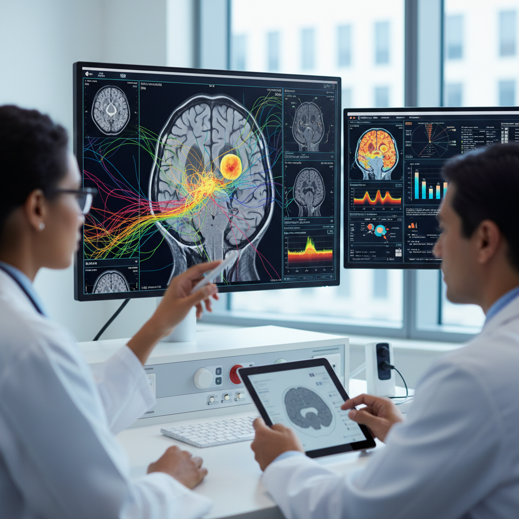
When it comes to brain cancer, imaging has never been just a picture. It helps answer some of the most important questions in care: where the tumour is, how far it has spread, whether it is growing, whether treatment is working, and in some cases whether what looks like progression might actually be an effect of therapy itself.
That is why advances in imaging are becoming one of the most important stories in modern neuro-oncology. The safest reading of the supplied evidence is that advanced imaging is improving brain tumour management by refining diagnosis, guiding treatment planning, and tracking response more precisely. Beyond that, emerging tools in radiomics and machine learning suggest that scans may begin to reveal clues about tumour biology itself, opening the door to more personalized care.
Caution still matters, though. The evidence package more clearly supports better decision-making than it does direct proof of major survival gains from imaging on its own.
MRI remains central — but it is no longer the whole story
Magnetic resonance imaging remains the clinical foundation of brain tumour assessment. It is still the main tool for identifying lesions, estimating their extent, defining critical anatomical relationships, and monitoring change over time.
But the picture is becoming more sophisticated. Advanced MRI methods, along with imaging such as PET, are adding information that goes beyond anatomy. Instead of only showing where a tumour is, they can also help show how it behaves.
That includes clues about:
- blood supply;
- metabolic activity;
- tissue composition;
- likely aggressiveness;
- and treatment response.
In practice, that matters because one of the biggest challenges in brain cancer care is that different tumours can look similar on standard scans, while the same tumour can change appearance over time or after treatment.
Seeing physiology and metabolism can improve the decision itself
The supplied evidence supports the idea that advanced MRI techniques and PET can provide physiological and metabolic information useful for:
- distinguishing tumour types;
- detecting small lesions;
- and assessing treatment response.
This is important because, in brain tumours, the real clinical challenge is often not simply spotting a mass. It is deciding whether a change on a scan reflects active tumour, biological evolution of disease, treatment-related inflammation, or therapy-induced tissue damage.
That distinction can alter major treatment decisions. It can influence whether a patient moves to surgery, radiotherapy, a chemotherapy change, close observation, or additional testing.
In other words, better imaging is not just prettier imaging. It is a way of making decisions with less uncertainty.
The next step is extracting information the human eye cannot easily see
That is where radiomics and machine learning come in. These approaches aim to extract quantitative patterns from images that are not obvious in a conventional visual read.
The logic is compelling: if a scan contains more information than the human eye can intuitively interpret, algorithms may help turn that hidden information into clinically useful markers.
That can include predictions about:
- tumour subtype;
- risk of progression;
- biological profile;
- expected response to treatment;
- and even immune-related behaviour.
A great deal of this still remains emerging rather than routine, but the direction is clear. Imaging is starting to move from being a visual confirmation tool to becoming a source of biomarkers.
One of the most promising examples comes from pediatric neuro-oncology
Among the supplied studies, one especially interesting signal comes from pediatric low-grade glioma. In that setting, multiparametric MRI combined with machine learning predicted immune profiles, prognosis, and treatment response.
That matters for two reasons. First, it suggests that imaging may help stratify risk more precisely. Second, it pushes imaging into a more active role in care: not only detecting and monitoring disease, but also guiding therapy decisions.
That is a meaningful conceptual shift. The scan is no longer just a diagnostic tool. It starts to participate in the reasoning about what kind of tumour is present in biological terms — and possibly which intervention makes the most sense.
This does not apply in exactly the same way to every brain tumour
At the same time, it is important not to overgeneralize.
The supplied evidence spans several tumour settings, including:
- glioblastoma;
- brain metastases;
- and pediatric low-grade glioma.
These are not biologically identical diseases, and they do not share the same treatment patterns or clinical questions. That means the usefulness of a given imaging technique in one context cannot simply be transferred to every other brain tumour setting.
This point matters because headlines about “advanced imaging” can sound universal. In practice, neuro-oncology still works by context, subtype, and the specific question being asked.
Imaging is becoming a bridge to precision neuro-oncology
Even with those differences, the evidence points to a broader trend: imaging is becoming an increasingly important part of precision neuro-oncology.
The wider glioblastoma literature supports the growing role of biomarker-informed care in brain cancer. In that movement, imaging may serve as a bridge between two clinical needs:
- understanding more about the tumour’s biology;
- and making practical decisions without always relying on repeated invasive procedures.
That may be one of the field’s biggest strengths. If imaging can function as a non-invasive biomarker, it could help personalize care more continuously over the course of illness.
What advanced imaging appears to improve right now
Even before the more futuristic promises, advanced imaging already seems to improve several practical parts of care, including:
- greater diagnostic precision;
- better surgical and radiotherapy planning;
- more refined response monitoring;
- and more careful interpretation of post-treatment changes.
That can help avoid major clinical errors, such as treating pseudoprogression as true treatment failure, or underestimating a small lesion with biologically important behaviour.
This is where the technology’s current value looks most solid.
What remains investigational
Despite the excitement, some of the most impressive advances are still investigational.
Machine-learning and radiomic tools still need:
- external validation;
- standardization across centres;
- integration into real clinical workflows;
- and consistent demonstration of usefulness outside highly specialized settings.
That means not every promising method is ready for broad routine use. In medicine, turning an elegant research finding into everyday practice is usually harder than it looks.
It is also important not to overstate imaging’s role on its own. It can help a great deal, but it usually works best as part of a larger framework that includes clinical assessment, pathology, molecular biology, and multidisciplinary judgment.
What this means for patients
For patients, the most useful message may be this: more advanced scans may make treatment more precise and better matched to the real behaviour of the tumour, even when they do not dramatically change everything.
That can mean a more reliable diagnosis, safer treatment planning, smarter monitoring, and less uncertainty when interpreting scans over time.
In the future, it may mean even more: imaging that helps anticipate risk, suggest biological subtype, and improve the timing or type of intervention. But that more ambitious future is still being built.
The balanced takeaway
The most responsible interpretation of the supplied evidence is that advanced imaging is becoming increasingly useful for diagnosing brain tumours more accurately, planning treatment, and tracking therapeutic response, while emerging radiomic and machine-learning tools point toward more personalized guidance.
MRI remains the clinical foundation, while advanced MRI and PET add physiological and metabolic information that may improve tumour distinction, reveal smaller lesions, and refine response assessment. In areas such as pediatric low-grade glioma, multiparametric imaging combined with machine learning already suggests more sophisticated uses in prognosis and risk stratification.
But the limits must stay clear: the implications are not identical across all brain tumours, some of the strongest advances remain investigational, and the best-supported message is improved decision-making rather than proof that imaging alone delivers major survival gains.
Even so, the direction matters. In brain cancer, treating better often begins with seeing better. Advanced imaging is doing exactly that — making the view more precise, more informative, and gradually more personalized.