New imaging tools are expanding how cancer is studied — but the evidence points more to a broader research leap than to a direct view inside living cells
New imaging tools are expanding how cancer is studied — but the evidence points more to a broader research leap than to a direct view inside living cells
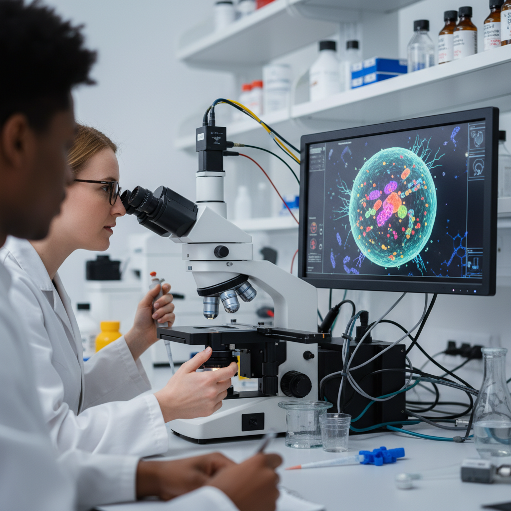
In cancer research, progress has always depended in part on learning how to see more clearly. Every new way of visualizing a tumour tends to change the kinds of questions scientists can ask. Where researchers once saw only a lump of abnormal tissue, they now try to distinguish microenvironments, cell interactions, metabolic patterns, heterogeneity, and the ways tumours adapt over time.
That is the backdrop to the new headline claiming that new imaging tools are helping cancer researchers “see inside living cells”. It is an arresting phrase, because it suggests almost direct access to the inner workings of cancer biology. But the most responsible reading of the supplied evidence requires some restraint. The references support a broader idea well: new imaging approaches and more realistic model systems are improving how cancer is studied. What they do not directly validate is one clearly established new routine method for observing intracellular processes in living cancer cells exactly as the headline suggests.
Seeing better has always changed oncology
Cancer has always been, in part, a visibility problem. First, it had to be seen in the body. Then it had to be examined under the microscope. Later, researchers learned to identify proteins, signalling pathways, mutations, and metabolic patterns. Today, the ambition is larger still: to understand the tumour as a dynamic system in which location, interaction, and function matter just as much as the visible lesion itself.
That matters because cancer is not simply a collection of rapidly growing cells. It is also a complex biological environment involving:
- different tumour cell populations;
- blood vessels;
- immune cells;
- extracellular matrix;
- regions with different oxygen and nutrient conditions;
- and uneven responses to treatment.
The better these layers can be visualized, the better researchers can understand what makes a tumour grow, resist treatment, or spread.
What the supplied evidence supports most clearly
Among the supplied references, the strongest support lies in the broader claim that advances in molecular imaging and three-dimensional cancer models are improving researchers’ ability to study tumour biology with more spatial and functional detail.
One important thread in the material involves FAP-based PET imaging. Fibroblast activation protein has become a useful target because it helps visualize components of tumour-associated stroma and other features of the cancer microenvironment. This kind of imaging does not simply reveal where a tumour is located. It can also provide clues about the biological activity surrounding it.
That is a meaningful advance. In many cancers, disease behaviour depends not only on malignant cells themselves, but also on the surrounding tissue and the interactions that support invasion, inflammation, and treatment resistance. Imaging methods that make that biology more visible widen the field of view for both researchers and, in some cases, clinicians.
The role of three-dimensional tumour models
Another important part of the supplied evidence is not strictly about imaging at all, but about three-dimensional cancer culture systems, which allow tumours to be studied in settings that more closely resemble real tissue than standard two-dimensional cultures.
That matters because tumour architecture changes behaviour. In 3D systems, researchers can better observe:
- tissue architecture;
- tumour heterogeneity;
- nutrient and oxygen gradients;
- cell-to-cell interactions;
- and drug responses in more realistic biological conditions.
These systems are not imaging tools in the narrow sense, but they act as essential companions to imaging. They create environments in which newer methods of visualization and analysis can capture biological behaviour that would be far harder to see in oversimplified laboratory systems.
What this really means: more context, not necessarily routine direct intracellular viewing
This is the key distinction. The supplied references support the idea that scientists are gaining a richer and more functional view of cancer biology. But that is not the same as proving that there is now a specific, validated method for directly seeing inside living cancer cells in routine use.
There are several reasons for caution.
First, FAP-based PET imaging is extremely useful, but it is not the same as intracellular live-cell imaging in the literal sense implied by the headline. It is a form of molecular and functional imaging at the tumour and microenvironment level, not a direct routine window onto fine intracellular processes inside living human cancer cells.
Second, the 3D culture literature is highly relevant to studying cancer more realistically, but it is not itself an imaging method. It is a model system that makes biological observation more meaningful.
Third, one of the supplied references is only indirectly relevant to the imaging claim, which reinforces the need to avoid drawing stronger conclusions than the evidence allows.
What the headline is probably trying to capture
Even with those limitations, the headline does reflect a real shift in cancer science. Research is moving towards combinations of:
- molecular imaging;
- spatial analysis;
- functional characterization;
- 3D tumour modelling;
- and more refined reading of the tumour microenvironment.
In practice, that means researchers are increasingly able to study cancer not just as a static target, but as a living system organized in space and shaped by multiple biological layers.
That is important because it changes the questions that can be asked. Instead of simply asking, “Where is the tumour?”, researchers can ask:
- which biological components are active;
- how the microenvironment contributes to disease behaviour;
- which regions appear more aggressive;
- and how tumours adapt under therapeutic pressure.
Why this matters for diagnosis and treatment strategy
Better imaging tools are not useful only because they produce more impressive images. They can influence how cancer is characterized and followed.
In molecular imaging, for example, the gains may include:
- more refined diagnosis;
- better assessment of disease extent;
- improved identification of therapeutic targets;
- stronger monitoring of treatment response;
- and, in some settings, tighter integration of diagnosis and treatment through theranostic approaches.
This is one of the strongest points in the newer PET-based approaches. They help connect tumour biology more directly to clinical decision-making than purely anatomical imaging often can.
What the story gets right
The story gets something important right by highlighting that newer tools are making cancer research more precise and more sophisticated. It is also right to suggest that visualizing a tumour now means more than simply locating a mass. It increasingly means trying to understand its biology in action.
It also points to a real trend: the boundaries between imaging, experimental modelling, and computational analysis are becoming less distinct. What allows researchers to “see better” today is often not one machine, but a combination of methods that makes cancer more observable across multiple scales.
What should not be overstated
At the same time, it would be too strong to use the supplied references to claim that a new technology has now been directly validated for broadly and routinely visualizing intracellular processes in living human cancer cells.
The safer reading is more limited:
- newer imaging tools are improving visibility into tumour biology;
- 3D systems are making cancer behaviour easier to study in realistic settings;
- molecular imaging such as FAP-PET is expanding access to tumour-associated biology;
- but that is not the same as showing, from the supplied evidence, that scientists now routinely “see inside living cells” in the strongest literal sense.
That distinction matters because, in science, a broad methodological advance should not be confused with a single established application already ready for routine use.
The real value of this kind of progress
Even with those cautions, the methodological advance still matters a great deal. Oncology depends heavily on the ability to connect structure, function, and biological context. Whenever a new tool improves that connection, it expands the potential for discovery.
Over time, that could mean:
- better understanding of tumour heterogeneity;
- more precise identification of therapeutic targets;
- experimental models that better reflect real tumours;
- and tighter integration between diagnosis, monitoring, and treatment.
That may be the most important point here: new tools do not need to literally show every intracellular event in order to transform research. They only need to make important parts of tumour biology more visible than they were before.
The most balanced reading
The supplied evidence supports a weak but coherent conclusion: new imaging tools and improved experimental systems are helping scientists study cancer with greater spatial, functional, and microenvironmental detail. FAP-based molecular imaging broadens the visibility of biologically relevant tumour features, while 3D cancer models make it easier to observe tumour architecture and interaction patterns in more realistic settings.
But the responsible interpretation has to recognize the limits. The supplied references do not directly validate one specific new method for seeing intracellular processes inside living cancer cells exactly as the headline implies. They more clearly support a broader improvement in the research toolkit than a single new established way of looking directly inside living cells.
The safest conclusion, then, is this: researchers are gaining much better tools for studying tumour biology in depth, and that is a real advance. But based on the material provided here, that progress is best understood as an important methodological leap in cancer research — not as a definitive new routine window into the inside of living cancer cells.